
Technical validation of CT Angiography in stroke management (CTA – Computed Tomography Angiography)
[pro_ad_display_adzone id=”17752″]
Disease and/or Condition: Acute stroke
Precision, Repeatability and Reproducibility of CT Angiography
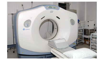
CT Angiography utilizes standard CT scanning techniques with the injection of iodine rich contrast material for diagnosing and evaluating vascular diseases and/or related conditions, such as blockages or aneurysms.[2] CTA has undoubted diagnostic and prognostic role in primary stroke imaging, as it enables the exploration, identification, and ruling out various anatomical and/or pathological processes related to the occlusion site, integrity of vessels, and collateral flow, all of which contribute to decision making in the urgency setting.
CT angiography is the device of choice for characterization of vascular anatomy to clear and locate the vessel occlusion site and to accurately identify the collateral status (identifying leptomeningeal collaterals, and determining the core expansion and the possibility of hemorrhagic transformation). Moreover, its influence on recanalization techniques is undoubted, as it can be used successfully to rule out the presence of calcifications and atherosclerosis.[1]
Patient Safety and Experience
- CTA necessitate exposition to iodinated contrast and radiation, which are potential risks of usage
- Obese patients may not fit into the opening of a conventional CT scanner, and/or their weight may be over the weight limit of the moving table (450 pounds).
- CT angiography to be avoided in patients with allergy to contrast material, in patients with advanced kidney disease or in those suffering of severe diabetes, as x-ray iodine-rich contrast material may be harmful.
Accuracy
- Allows the exploration and ruling out the site of vascular occlusion, as well as it allows ruling out the existence of calcifications and atherosclerosis, that affect recanalization techniques positively
- It is the optimal noninvasive imaging device to explore leptomeningeal collaterals, which not only define the ratio of core protraction, but also show the potency of hemorrhagic outbreak, where patients with poor collateral status are at a higher risk of intra-cranial hemorrhagic transformation [3]
- Having the ability to show a bad collateral-score, it undoubtedly assists in discovering a detrimental profile (larger DWI baseline lesion volume – higher median NIHSS and functional reliance in a 3 months post-stroke period) [4].
- CT Angiography aid physicians by providing and backing them up with more precise images of your blood vessels that no MRI can provide [5]
Availability
- CTA is available in most hospitals and healthcare centers with stroke services.
References
- emc.healthyorthodoxmedicine
- radiologyinfo.org/en/info.cfm?pg=angioct
- A.M. Demchuk, F. Khan, M. D. Hill et al., “Importance of leukoaraiosis on CT for tissue plasminogen activator decision making: evaluation of the NINDS rt-PA stroke study,” Cerebrovascular Diseases, vol. 26, no. 2, pp. 120–125, 2008
- L.C. Souza, A. J. Yoo, Z. A. Chaudhry, et al., “Malignant CTA collateral profile is highly specific for large admission DWI infarct core and poor outcome in acute stroke,” American Journal of Neuroradiology, vol. 33, no. 7, pp. 1331–1336, 2012
- radiopaedia.org/articles/ct-perfusion-in-ischaemic-stroke
Verified by: Dr.Diab (January 18, 2017)
Citation: Dr.Diab. (January 18, 2017). Technical validation of CT Angiography in stroke management. Medcoi Journal of Medicine, 3(2). urn:medcoi:article17250.


There are no comments yet
Or use one of these social networks