
Imaging biomarkers definition modalities and classification
[pro_ad_display_adzone id=”17752″]
What is a biomarker
A biomarker is a measurable entity that can be used as an indicator of normal functional biological processes, or as a response to a therapeutic intervention [1][2]. They are often used along the course of a therapy for the diagnosis, prognosis or monitoring of the clinical response of the organism to therapy, it can be classified into imaging biomarkers such as CT or computed tomography, positron emission tomography, MRI or magnetic resonance imaging) or molecular biomarkers that can be used to refer to non imaging biomarkers owing biophysical properties.[3]
A biomarker is an authentic and previously validated and globally accepted disease characteristic that can be reliably and repeatedly measured in a cost effective and generalized manner, and which acts as a meaningful surrogate for pathological presence, absence, development and activity, or prognosis in individuals or groups with the disease process.
The mentioned above definitions include in vitro diagnostic measurements made from:
- Biologic fluids such as utilizing low-density lipoprotein cholesterol…
- Solid tissue pathologic specimens, biopsies and smears such as tumor biopsy, cervical smear, skin biopsy…
- Physiologic and biophysical measurements such as electrocardiographic data, blood pressure, etc…
- Measurements from images such as tumor size, tumor pharmacokinetics, rheumatoid erosions…
Imaging biomarkers
Imaging biomarkers can help detect and treat diseases earlier and more effectively, they also potentially reduce the financial cost pressuring healthcare providers today. Medical imaging could have a great impact on progressive diseases, such as certain types of cancer, atherosclerotic cardiovascular diseases, Alzheimer’s disease and rheumatoid arthritis.
They involve imaging technologies, characterized as noninvasive procedures that have many advantages, they are comfortable to patients and often produce intuitive and multidimensional results possessing both qualitative and quantitative data, and when combined with other sources of information can be very useful for doctor along the diagnostic procedures, as well as imaging biomarkers can be used to trace substances in vivo and identify their metabolites of leftovers in the body as in the pharmacokinetics assay.
Imaging biomarkers utilize traditional and genetic information to help physicians and medical practitioners characterize various pathological disorders such as the evaluation of tumors progression to rule out treatment effectiveness.
Biomarkers, however, require a substantial amount of supporting data to become a widely accepted surrogate clinical endpoint. However, some lab based tests have already reached this status:
- LDL cholesterol for identifying risk of stroke or myocardial infarction
- HgA1c for diabetes mellitus
- CD4/viral copy number for HIV
- Blood pressure for cardiovascular disease
Quantitative imaging includes the development, optimization and standardization, of molecular, functional, and anatomical imaging acquisition protocols, display methods, data analyses and reporting structures. These features allow the precise and accurate validation of obtained image-derived metrics with anatomical and physiological relevant parameters, including the prognosis and treatment response, permitting the utilization of such metrics in research and patient care.
According to the Toward Quantitative Imaging task force of the Radiological Society of North America (RSNA) quantitative imaging is the identification of extracted quantifiable features from medical images for the assessment of the status of a disease (the severity, degree of change, injury, etc…), whether the condition is chronic or relative to normal.[4]
Emerging imaging technologies
Imaging modalities:
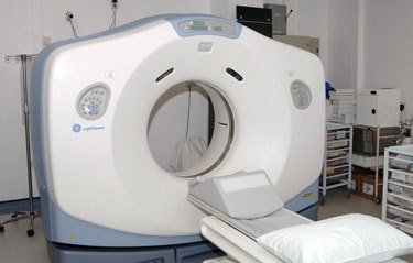
Computerized axial tomography (CT), Magnetic Resonance Imaging (MRI), positron emission tomography (PET), and nuclear imaging are already widely used in mainstream imaging and are now expanding into new dimensions.
Functional imaging advances provide an understanding of metabolism inside of the organism such as the metabolism of a tumor and its biological and anatomical pathways by viewing vascular permeability and blood flow allowing clinicians or physicians to view biological activity at the very cellular level (molecular base).
Imaging modalities can help in monitoring the marketing process (drug distribution), pharmacodynamics and pharmacokinetics essential for clinical trials conducted early in its lifecycle.
Examples:
F-PET and F-MRI provide a deeper molecular dimension.
- Dynamic imaging monitor molecular or systemic changes over time such as: Heart beating, profiles of receptors being blocked or unveiled over a dosing period of time.
- Structural imaging explores dimensions, volume and density ex. dimensions of the tumor (tumor size) which are valuable in understanding tumor growth (invisibility) and response, as well as monitoring the tumor volume, tumor edges and density.
Classification
Biomarkers can be classified as follows:
- Diagnostic (Disease related biomarkers which gives an indication of an already existing disease, its measurement improves the accuracy of patient diagnosis or prognosis)
- Predictive (shows the probable effect of a treatment in a patient, its measurement predicts which treatment would be suitable, it is recommended when selecting the therapy and for understanding whether the therapy is beneficial or not)
- Response (change in biomarker after initiating the treatment, it predicts whether the treatment is beneficial i.e. will lead to beneficial outcome, or not)
- Prognostic (may show the outcome of a diseases and how the disease may develop in a patient regardless of the treatment course).
- Monitoring (regular measurement of quantitative imaging biomarker to detect disease relapse or emergence of drug toxicity).
References
1- emc.healthyorthodoxmedicine
2- pubs.rsna.org/doi/full/10.1148/radiol.10100800
3- K. Hajjar, D. M. Kerr, and K. R. Lees, “Thrombolysis for acute ischemic stroke,” Journal of Vascular Surgery, vol. 54, no. 3, pp. 901–907, 2011.
4- Toward Quantitative Imaging (TQI) Workshop. RSNA Headquarters, Oak Brook, IL. July 10–11, 2008. Radiological Society of North America. //rsna.org/Research/TQI/upload/Workshop-Summary-FINAL.pdf. Accessed March 17, 2010
Verified by: Dr.Diab (January 18, 2017)
Citation: Dr.Diab. (January 18, 2017). Imaging biomarkers definition modalities and classification. Medcoi Journal of Medicine, 5(2). urn:medcoi:article17217.


There are no comments yet
Or use one of these social networks