
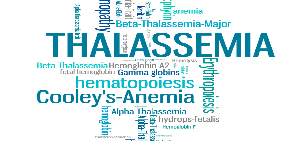
Thalassemia
Thalassemia is a common genetic hemolytic disorder characterized by defective hemoglobin Hb synthesis and ineffective erythropoiesis, the disease is common in mideast-southeast africa (α Thalassemia 25%, clinics show in 10% of patients).
Causes
The defect in Hb synthesis is caused by one decreased globin polypeptide chain (β, α, γ, δ)
β Thalassemia have two forms:
- Thalassemia minor characterized by mild microcytic anemia, characteristic in heterozygote carriers
- Thalassemia major, characteristic in homozygotes
α Thalassemia require two pairs of genes, it is classified into three forms:
- α Thalassemia silent carrier (2), no evidence of a clinical abnormality is seen, characteristic in heterozygotes of a single gene.
- α Thalassemia trait (1), it presents with a clinical picture identical to β Thalassemia minor, it is characteristic in heterozygotes of a double gene.
- Lethal form of α Thalassemia because Hb doesn’t transport O2, characteristic in homozygotes, homozygosity of both genes.
α chain deficiency results in increased β chains (HbH), and increased γ chains (barts Hb) in infancy.
Symptoms and Clinical picture
β Thalassemia minor is asymptomatic
β Thalassemia major (cooley’s anemia) is characterized by symptoms of severe anemia, chronic fatigue, expanded marrow space, Fe overload, jaundice, cholelithiasis (like in sickle cell anemia), leg ulcers and splenomegaly (enlarged spleen).
Bone marrow hyperactivity results in the thickening of the bones (cranial and malar eminence), with long bone involvement that is characterized by common pathological fractures.
Growth impairment, and puberty is either delayed or absent.
Fe deposits in the heart muscles, resulting in cardiac dysfunction and heart failure.
Hepatic siderosis results in cirrhosis.
Thalassemia minor (HbH disease) is characterized by hemolytic anemia and splenomegaly.
Diagnosis and Lab studies
serum bilirubin, serum Fe, and serum ferritin all increased.
Erythroid hyperplasia is noted in the bone marrow.
β-α Thalassemia minor, mild to moderate microcytic anemia, serum ferritin is checked to rule out Fe deficiency.
β Thalassemia major, severe anemia, Hb less or equal to 6g/dl, RBC elevated.
Blood smear: nucleated erythroblasts, target cells, small pale RBC, punctate and diffuse basophilia.
Quantitative Hb studies for diagnosis: increased HbA2 is characteristic of β Thalassemia minor. If HbA2 > 3% it is characteristic of β Thalassemia major. Whereas an elevated level of 90% HbF or more is characteristic of β Thalassemia major
In α Thalassemia HbA2 and HbF are normal.
HbH disease is diagnosed by fast migrating of HbH or Barts fraction on Hb electrophoresis.
DNA polymerase chain reaction for prenatal diagnosis.
In case of β Thalassemia major, x-ray shows thinned cortices of the skull and long bones, marrow space is widened.
Treatment
For children with β Thalassemia major, transfusion with Fe removal via chronic Fe-chelation therapy, as a precaution of Fe overload.
Splenectomy (total) to cure of hemosiderosis (increased deposition of Fe in tissues, especially in the lungs and kidneys).
Phlebotomy is suggested if the Fe level in the body > 5g (normally 2.5g for females and 3g for males), as it can result in hemochromatosis where Fe levels can reach up to 50g…
Verified by: Dr.Diab (January 17, 2017)
Citation: Dr.Diab. (January 17, 2017). Thalassemia causes symptoms and treatments. Medcoi Journal of Medicine, 7(2). urn:medcoi:article16168.


There are no comments yet
Or use one of these social networks